Normal Clavicle X Ray

Normal Clavicle X Ray

Fractures of the clavicle are common, up to 10% of all fractures. The mechanism of injury is usually medium to high energy falling on an outstrecthed arm, in direct impact sports. The joints at the ends of the bone, where it connects to the breastbone and. Los rayos x de la clavcula son una tcnica de imagenologa utilizada para examinar la clavcula, un hueso largo y delgado que se encuentra entre el esternn y el hombro. Because of its ligamentous attachments and the presence of articulations at both ends, the clavicle can also be involved in arthritic diseases.

Fractures of the clavicle are common, up to 10% of all fractures. The mechanism of injury is usually medium to high energy falling on an outstrecthed arm, in direct impact sports. The joints at the ends of the bone, where it connects to the breastbone and. Los rayos x de la clavcula son una tcnica de imagenologa utilizada para examinar la clavcula, un hueso largo y delgado que se encuentra entre el esternn y el hombro. Because of its ligamentous attachments and the presence of articulations at both ends, the clavicle can also be involved in arthritic diseases.

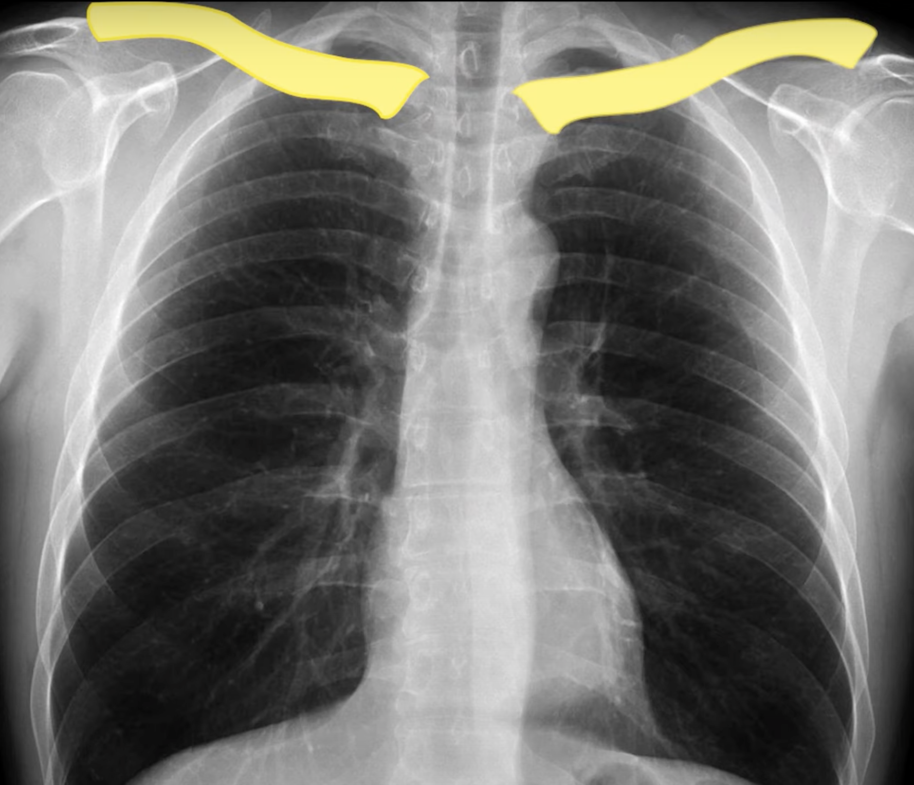
An apical or lordotic view may then provide. Normal clavicle radiographs showing the two standard projections. Uq radiologic anatomy 7. Upper limb 7. 1 shoulder & clavicle.

Elwyn Davidson School Calendar 2024-2025

Mark Consuelos Salary On Live

Can Chickens Eat Almond Flour

Chest X-Ray For Students: How to Interpret and Present Methodically
Acromio clavicular seperation | Image | Radiopaedia.org
acromio clavicular radiopaedia joint seperation radiology injuries acromioclavicular injury version size full
X-ray Image of Clavicle, AP View Stock Image - Image of black, accident
clavicle ap ray preview